Google and Harvard scientists create 3D map of the human brain
غوغل وهارفارد يطوران أكثر الخرائط ثلاثية الأبعاد تفصيلا للدماغ البشري
News Agencies
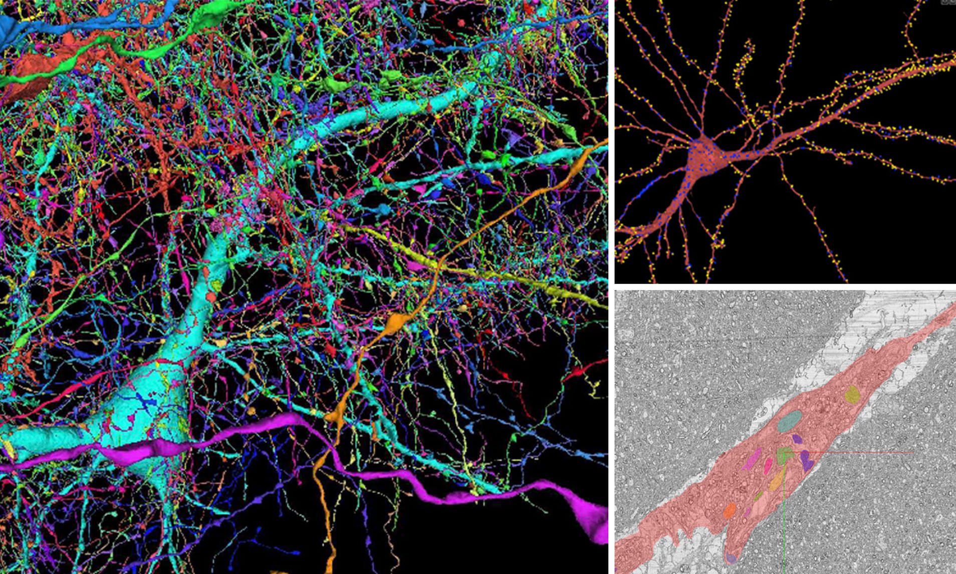
Demonstrating just how extraordinary of an organ the human brain is, a team of scientists with Google and Harvard University has just conjured up an amazing color-coded map of nearly 4,000 incoming axons connecting to a single neuron.
This stunning browsable 3D map which represents just one millionth of the cerebral cortex has been composed using 225 million separate images and a super computer-crunching 1.4 petabytes of data. The main goals of this project were to produce a novel resource for studying human brains and to improve and scale the underlying connectomics technologies.
Mapping the convoluted clutter of neurons, synapses and neurological cells is a formidable task at best, but these diligent engineers achieved admirable results with this ambitious grey-matter project to create some sort of human brain wiring diagram (“connectome”) based on its 86 billion neurons linked via 100 trillion synapses.
Back in January 2020, Google released the fly “hemibrain” connectome, an online database delivering the morphological structure and synaptic connectivity of approximately half of a fruit fly brain. This detailed database and its visualization tools has totally altered the way that neural circuits and pathways are understood in the fly brain.
Now in a huge advancement, Google and the Lichtman Lab at Harvard University has presented a mind-boggling model of a micro section of the human brain, which began with a microscopic sample obtained from the temporal lobe of a human cerebral cortex. This slice was stained for added visual clarity, then coated in resin to protect and preserve it.
The surgical sample was then diced into roughly 5,300 slices each around 30 nanometers (nm) thick. A scanning electron microscope was employed next to image the sample, with a maximum resolution registering down to 4 nm. This all resulted in the creation of 225 million two-dimensional images that could be stitched together to manifest a single 3D volume.
Machine learning algorithms carefully scanned the slice to identify the different cells and their underlying structures. Following a few passes by various automated systems, human eyes provided a visual edit pass for some cells to determine whether or not the algorithms were identifying them correctly.
Google has named this result the H01 dataset, and it’s considered to be perhaps the most comprehensive maps of the human brain in existence. Containing 50,000 cells and 130 million synapses, in addition to tinier cellular segments like axons, dendrites, myelin, and cilia, this startling rendering was born only by utilizing over a million gigabytes of data.
But the insane element to this advancement is that Google labels this miniature sample as just one millionth of the volume of the entire human brain.
As the Lichtman Lab team scales this behemoth brain map even further in the future, the whole H01 dataset is available online for researchers and scholars to investigate, with a preprint paper showing the work available on bioRxiv.
قنا
واشنطن: طورت شركة /غوغل/ الأمريكية بالتعاون مع علماء في جامعة /هارفارد/، خريطة ثلاثية الأبعاد لجزء صغير من دماغ الإنسان، باستخدام (225) مليون صورة وأكثر من مليون غيغابايت من البيانات.
وتعد الخريطة ثلاثية الأبعاد أول دراسة واسعة النطاق للتوصيل المشبكي في القشرة الدماغية البشرية التي تمتد على أنواع متعددة من الخلايا عبر جميع طبقات القشرة الست.
وتُظهر الخريطة المشفرة بالألوان 4000 محور عصبي متصل بخلية عصبية واحدة، وهي الخلية الأساسية في النظام العصبي للإنسان، والمسؤولة عن تلقي المدخلات الحسية، وعلى الرغم من أنها لا تمثل سوى ملليمتر مكعب واحد من أنسجة المخ، إلا أن هذه هي أكبر عينة من هذه الأنسجة صُوّرت وأعيد بناؤها بهذه التفاصيل الدقيقة، وتشمل عشرات الآلاف من الخلايا العصبية، وملايين شظايا الخلايا العصبية، و130 مليون من نقاط الاشتباك العصبي والتركيبات تحت الخلوية، بما في ذلك المحاور والأهداب والتشعبات.
وقالت /غوغل/ إن الأهداف الرئيسية للمشروع كانت إنتاج طريقة جديدة لدراسة الدماغ البشري مع تحسين التكنولوجيا الأساسية المستخدمة في رسم خريطة للجهاز العصبي للكائن الحي.
ولإنشاء الخريطة، أخذ الباحثون في غوغل ومختبر ليتشمان بالجامعة، عينة مجهرية من الفص الصدغي لقشرة دماغية بشرية، وكانت عينة الأنسجة هذه مغلفة بمادة صمغية، قبل تقطيعها إلى 5300 شريحة، يبلغ سمك كل منها حوالي 30 نانومتر، وقام مجهر إلكتروني يمكنه التكبير إلى حوالي 4 نانومتر، بفحص العينة، ما أدى إلى إنشاء 225 مليون صورة ثنائية الأبعاد “يمكن تجميعها معا لإظهار حجم واحد ثلاثي الأبعاد.
وتعد القشرة الدماغية هي الجزء الأكبر من دماغ الثدييات، وغالبا ما يُطلق عليها اسم “المادة الرمادية”، مع ما يصل إلى ست طبقات من الخلايا العصبية مسؤولة عن العديد من الوظائف مثل التفكير والتخطيط والإدراك واللغة والذاكرة والحركة الإرادية.
وقُدّمت عينات الأنسجة الخاصة بالمشروع من قبل المرضى في مستشفى /ماساتشوستس/ العام، حيث يقوم الجراحون بإزالة أجزاء من القشرة الدماغية الطبيعية أثناء إجراء الجراحة للتخفيف من الصرع.


