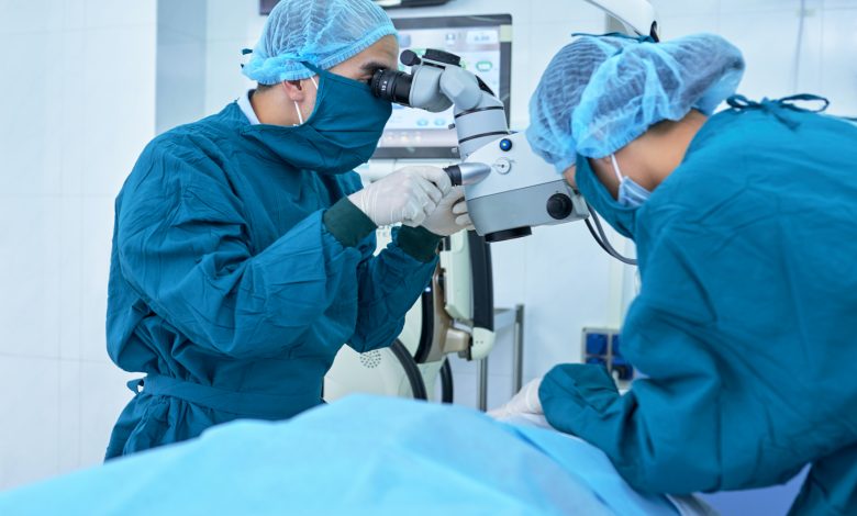
HMC performs surgery to remove a brain tumor on a patient while awake
حمد الطبية تجري جراحة لإزالة ورم دماغي لمريضة في وضع اليقظة
Al-Raya Medical – WGOQatar Translations
Doha: Doctors at Hamad Medical Corporation performed complex and precise surgery on a 55-year-old patient while awake, making it the first successful surgery in Qatar using cortical brain mapping technology.
Dr. Sirajuddin Balkheir, Head of Neurosurgery and Director of The Residency Program for Neurosurgery at Hamad Medical Corporation, said the surgery , known as the “cranial pilgrimage”, was performed on a patient with a diffuse brain tumor, where cortical stimulation planning technique was used to identify an important area of the brain.

He pointed out that this type of brain surgery takes place for the patient in a position of alertness and attention, where neurosurgeons and the brain electricity team perform surgery known as cortical stimulation planning, so that the brain is exposed and during the operation the surface of the brain is stimulated using a small electric probe to treat nerve problems of the brain such as tumors, epileptic seizures and vascular abnormalities in the brain.
Dr. Sirajuddin explained that brain surgery in alert mode allows surgeons to ask patients to perform some motion tests along with reading and talking while the open brain is stimulated so that they can determine the safest way to reach the tumor.

He added that at the beginning of the surgery, during the incision of the skin and the removal of the skull, the patient was given a small dose of the anesthetic by the anesthesia team and then was awakened after opening the brain where the patient was talking during the operation and asked to move her upper and lower limbs to ensure the maintenance of her functions as a special system was used to accurately locate the tumor in the patient’s brain, explaining that the performance of the brain during the surgeon’s operation is also monitored to ensure the safety of the brain during the removal of the tumor.

He explained that the surgery took less than three hours, followed the next day by an MRI scan where it showed the complete removal of the brain tumor, adding that the patient was able to leave the hospital two days after the operation, pointing out that the ability to remove the tumor using this technique enables patients to recover faster, which improves the quality of life by reducing the chances of complications caused by the operation.
For his part, Dr. Abdullah Al Ansari, Chief Medical Officer of Hamad Medical Corporation commented on the success of the operation, describing it as an important achievement for the neurosurgery team and Hamad Medical Corporation, explaining that work is constantly being done to bring the best technologies to the population in Qatar, and to develop the quality of health care, adding that the ability of the qualified neurosurgeon team to successfully deliver this surgery in Qatar for the first time means that patients do not need to travel to get this kind of specialized care.
الراية الطبية
الدوحة: أجرى أطباء بمؤسسة حمد الطبية عملية جراحية مُعقدة ودقيقة لمريضة تبلغ من العمر 55 عامًا، وهي في وضع اليقظة، ما يجعلها أول عملية جراحية تجرى بنجاح في دولة قطر باستخدام تقنية خرائط الدماغ القشرية.
وقال الدكتور سراج الدين بالخير، رئيس قسم جراحة المخ والأعصاب ومدير برنامج الإقامة لجراحة المخ والأعصاب في مؤسسة حمد الطبية إنه تم إجراء الجراحة – المعروفة باسم «حج القحف» – لمريضة تعاني من إصابة وورم مُنتشر في الدماغ، حيث تم استخدام تقنية تخطيط التحفيز القشري لتحديد منطقة مهمة في الدماغ.

ولفت إلى أن هذا النوع من جراحة الدماغ يجري للمريض وهو في وضع اليقظة والانتباه، حيث يُجري جراحو الأعصاب وفريق الكهرباء الدماغية جراحة تعرف باسم تخطيط التحفيز القشري، بحيث يكون الدماغ مكشوفًا ويتم خلال العملية تحفيز سطح الدماغ باستخدام مسبار كهربائي ضئيل الحجم بحيث يعمل على علاج المشاكل العصبية للدماغ كالأورام ونوبات الصرع وتشوهات الأوعية الدموية في الدماغ.
وأوضح الدكتور سراج الدين، أن جراحة الدماغ في وضع اليقظة تسمح للجرّاحين الطلب من المرضى القيام ببعض اختبارات الحركة إلى جانب القراءة والحديث بينما يتم تحفيز الدماغ المفتوح ما يُمكّنهم من تحديد الطريق الأكثر أمانًا للوصول إلى الورم.

وأضاف أنه في بداية العملية، وأثناء شق الجلد وإزالة الجمجمة، تم إعطاء المريضة جُرعة صغيرة من المُخدر من قِبل فريق التخدير وبعدها تم إيقاظها بعد فتح الدماغ حيث كان يتم التحدث إلى المريضة أثناء إجراء العملية وطلب منها تحريك أطرافها العلوية والسفلية للتأكد من الحفاظ على وظائفها كما تم استخدام نظام خاص لتحديد مكان الورم بدقة في دماغ المريضة، مُوضحاً أنه تتم أيضًا مُراقبة أداء الدماغ خلال إجراء الجراح للعملية بما يضمن الحفاظ على سلامة الدماغ أثناء إزالة الورم.
وأوضح أن الجراحة استغرقت أقل من ثلاث ساعات، وتبعها في اليوم التالي إجراء فحص بالرنين المغناطيسي حيث أظهر الإزالة التامة للورم الدماغي، مضيفًا أن المريضة استطاعت مُغادرة المستشفى بعد يومين من إجراء العملية، لافتًا إلى أن القدرة على إزالة الورم باستخدام هذه التقنية يُمكّن المرضى من التعافي بصورة أسرع مما يُحسّن في جودة الحياة من خلال التقليل من فرص حدوث مُضاعفات ناجمة عن العملية.

من جهته علّق الدكتور عبدالله الأنصاري، الرئيس الطبي بمؤسسة حمد الطبية على نجاح العملية واصفًا إياها بالإنجاز الهام لفريق جراحة الأعصاب ولمؤسسة حمد الطبية، موضحًا أنه يتم العمل باستمرار لجلب أفضل التقنيات للسكان في دولة قطر، ولتطوير جودة الرعاية الصحيّة، مضيفًا أن قدرة فريق جراحة الأعصاب المُؤهل على تقديم هذه الجراحة بنجاح في دولة قطر للمرة الأولى، يعني عدم حاجة المرضى للسفر للحصول على مثل هذا النوع من الرعاية المتخصصة.


